Total hip arthroplasty
The hip joint is one of the largest and most important joints in the body. The hip joint is one of the most mobile joints in the body. The head of the femur (also known as the femur) is in the shape of a round sphere and is completely surrounded by the acetabular cavity, which is in the shape of a hollow bowl. The curvature of this ball and the bowl are perfectly coordinated with each other, so that the ball or sphere can easily move in any direction inside the bowl, and this causes the hip joint to have a wide range of motion in different directions. However, the hip joint is very stable.
Both the upper surface of the head of the femur and the inner surface of the acetabulum cavity are covered with a white and elastic layer called cartilage, whose function is to make it slippery and reduce the friction between these two articular surfaces, thus facilitating the movement of the joint. This cartilaginous layer causes arthritis and wear of the hip joint.

The hip joint is surrounded and enclosed by the joint capsule. The joint capsule creates a closed space inside which the head of the femur and the cavity of the acetabulum are located. A thin layer called synovial membrane covers the inner surface of the hip joint capsule and its function is to secrete a viscous liquid called joint fluid or synovial fluid. This liquid makes joint movement smooth (similar to lubrication in cars) and also nourishes joint cartilage.
Description of surgery
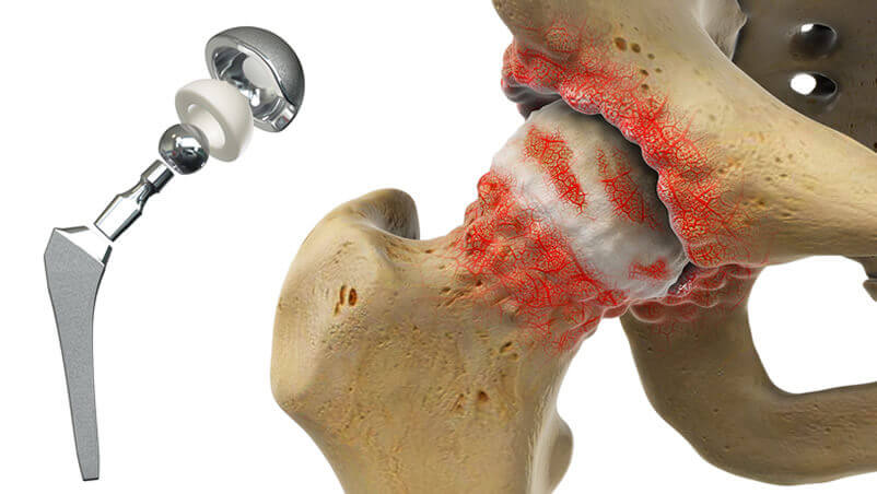
In hip replacement surgery or total joint arthroplasty, the damaged bone and cartilage are removed and replaced with prosthetic components. He removes the damaged femur head from the patient's body and replaces it with a metal ball. This metal head is connected to the remaining femur with an appendage called the Stem handle, which is connected to the metal head and by placing the handle inside The central canal of the artificial femoral head is secured in place.
The orthopedic surgeon then scrapes the inner surface of the acetabulum cavity in the hip area, which is damaged, with special tools. With this, the damaged cartilages and extra bones are removed from the acetabular cavity and the cavity becomes a complete hemisphere. Then a metal cup in the shape of a hemisphere is placed inside the prepared cavity and a poly plastic bowl is placed inside it. Ethylene is placed.

In some cases, the plastic cup may be attached to the acetabular cavity without a metal cup and only by a special adhesive. That glue is called bone cement. The metal head is placed inside the plastic bowl so that it can move in different directions inside it.